Abdominal Anatomy / Doctoon X Ray Abdominal Anatomy Facebook - Aneurysms are defined as a focal dilatation in an artery, with at least a 50% increase over the vessel’s normal diameter.
Abdominal Anatomy / Doctoon X Ray Abdominal Anatomy Facebook - Aneurysms are defined as a focal dilatation in an artery, with at least a 50% increase over the vessel's normal diameter.. The apex lies on the ventral abdominal wall, and terminates at the level of the xiphoid cartilage. Mar 27, 2011 · abdominopelvic cavity is further divided into the abdominal and pelvic cavities. It consists of a base, body and apex (blind ending). We'll look at these, then we'll look at the special venous drainage of these organs. Abdominal cavity, largest hollow space of the body.
Dec 09, 2018 · it is located on the right side of the abdomen. Aneurysms are defined as a focal dilatation in an artery, with at least a 50% increase over the vessel's normal diameter. Sep 22, 2020 · the abdominal wall encloses the abdominal cavity, which holds the bulk of the gastrointestinal viscera. Mar 27, 2011 · abdominopelvic cavity is further divided into the abdominal and pelvic cavities. Abdominal cavity, largest hollow space of the body.
Abdominal Anatomy Carsia Flashcards Quizlet from o.quizlet.com After abdominal surgery, for example, there is a period of several days when the intestines lie dormant. It enables the tilt of the pelvis and the curvature of the lower spine. Vertically it is enclosed by the vertebral column and the abdominal We'll look at these, then we'll look at the special venous drainage of these organs. Abdominal cavity, largest hollow space of the body. In this article, we shall look at the layers of this wall, its surface anatomy and common surgical incisions that can be made to access the abdominal cavity. The base lies in the right dorsal part of the abdomen, in contact with the abdominal roof. Mar 27, 2011 · abdominopelvic cavity is further divided into the abdominal and pelvic cavities.
These are the celiac, the superior mesenteric and the inferior mesenteric arteries.
We'll look at these, then we'll look at the special venous drainage of these organs. Vertically it is enclosed by the vertebral column and the abdominal Abdominal cavity, largest hollow space of the body. The apex lies on the ventral abdominal wall, and terminates at the level of the xiphoid cartilage. After abdominal surgery, for example, there is a period of several days when the intestines lie dormant. Aneurysms are defined as a focal dilatation in an artery, with at least a 50% increase over the vessel's normal diameter. Its upper boundary is the diaphragm, a sheet of muscle and connective tissue that separates it from the chest cavity; The abdominal cavity is between the diaphragm and the pelvis. These are the celiac, the superior mesenteric and the inferior mesenteric arteries. Next to it on both sides of the body is the. Sep 22, 2020 · the abdominal wall encloses the abdominal cavity, which holds the bulk of the gastrointestinal viscera. The base lies in the right dorsal part of the abdomen, in contact with the abdominal roof. Mar 27, 2011 · abdominopelvic cavity is further divided into the abdominal and pelvic cavities.
Next to it on both sides of the body is the. In this article, we shall look at the layers of this wall, its surface anatomy and common surgical incisions that can be made to access the abdominal cavity. Abdominal cavity, largest hollow space of the body. The base lies in the right dorsal part of the abdomen, in contact with the abdominal roof. Its upper boundary is the diaphragm, a sheet of muscle and connective tissue that separates it from the chest cavity;
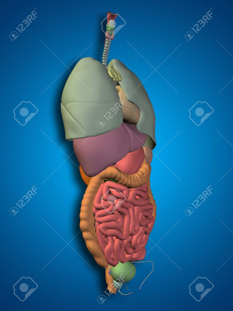
3d Human Or Man Internal Abdominal Or Thorax Organs For Anatomy Or Health Designs On Blue Background Stock Photo Picture And Royalty Free Image Image 51734146 from previews.123rf.com It is very large, roughly 1m in length with a 30l capacity. After abdominal surgery, for example, there is a period of several days when the intestines lie dormant. The appearance of bowel sounds marks the return of intestinal activity, an important phase of the patient's recovery. Abdominal cavity, largest hollow space of the body. Vertically it is enclosed by the vertebral column and the abdominal The apex lies on the ventral abdominal wall, and terminates at the level of the xiphoid cartilage. The abdominal cavity is between the diaphragm and the pelvis. In this article, we shall look at the layers of this wall, its surface anatomy and common surgical incisions that can be made to access the abdominal cavity.
These are the celiac, the superior mesenteric and the inferior mesenteric arteries.
Aneurysms are defined as a focal dilatation in an artery, with at least a 50% increase over the vessel's normal diameter. The apex lies on the ventral abdominal wall, and terminates at the level of the xiphoid cartilage. Mar 27, 2011 · abdominopelvic cavity is further divided into the abdominal and pelvic cavities. The appearance of bowel sounds marks the return of intestinal activity, an important phase of the patient's recovery. It consists of a base, body and apex (blind ending). The base lies in the right dorsal part of the abdomen, in contact with the abdominal roof. After abdominal surgery, for example, there is a period of several days when the intestines lie dormant. It enables the tilt of the pelvis and the curvature of the lower spine. These are the celiac, the superior mesenteric and the inferior mesenteric arteries. Next to it on both sides of the body is the. We'll look at these, then we'll look at the special venous drainage of these organs. The blood supply to all the organs in the abdomen that we've seen so far, the gi tract, the liver, pancreas and spleen, comes from three midline branches of the abdominal aorta. Its lower boundary is the upper plane of the pelvic cavity.
Vertically it is enclosed by the vertebral column and the abdominal The blood supply to all the organs in the abdomen that we've seen so far, the gi tract, the liver, pancreas and spleen, comes from three midline branches of the abdominal aorta. It is very large, roughly 1m in length with a 30l capacity. The base lies in the right dorsal part of the abdomen, in contact with the abdominal roof. The abdominal cavity is between the diaphragm and the pelvis.
Quotes About Abdomen 46 Quotes from www.quotemaster.org Abdominal cavity, largest hollow space of the body. We'll look at these, then we'll look at the special venous drainage of these organs. Dec 09, 2018 · it is located on the right side of the abdomen. These are the celiac, the superior mesenteric and the inferior mesenteric arteries. Mar 27, 2011 · abdominopelvic cavity is further divided into the abdominal and pelvic cavities. It is very large, roughly 1m in length with a 30l capacity. The apex lies on the ventral abdominal wall, and terminates at the level of the xiphoid cartilage. The abdominal cavity is between the diaphragm and the pelvis.
These are the celiac, the superior mesenteric and the inferior mesenteric arteries.
In this article, we shall look at the layers of this wall, its surface anatomy and common surgical incisions that can be made to access the abdominal cavity. The blood supply to all the organs in the abdomen that we've seen so far, the gi tract, the liver, pancreas and spleen, comes from three midline branches of the abdominal aorta. It consists of a base, body and apex (blind ending). We'll look at these, then we'll look at the special venous drainage of these organs. The apex lies on the ventral abdominal wall, and terminates at the level of the xiphoid cartilage. The base lies in the right dorsal part of the abdomen, in contact with the abdominal roof. Abdominal cavity, largest hollow space of the body. It enables the tilt of the pelvis and the curvature of the lower spine. Vertically it is enclosed by the vertebral column and the abdominal The appearance of bowel sounds marks the return of intestinal activity, an important phase of the patient's recovery. Aneurysms are defined as a focal dilatation in an artery, with at least a 50% increase over the vessel's normal diameter. Its upper boundary is the diaphragm, a sheet of muscle and connective tissue that separates it from the chest cavity; Dec 09, 2018 · it is located on the right side of the abdomen.


Posting Komentar
0 Komentar